It is such a privilege to practice a profession that can save vision, relieve debilitating symptoms and protect the future of my patients’ vision. I thought it might be a good time to have a retrospective look at some of these amazing advances over my career.
Retinal Imaging
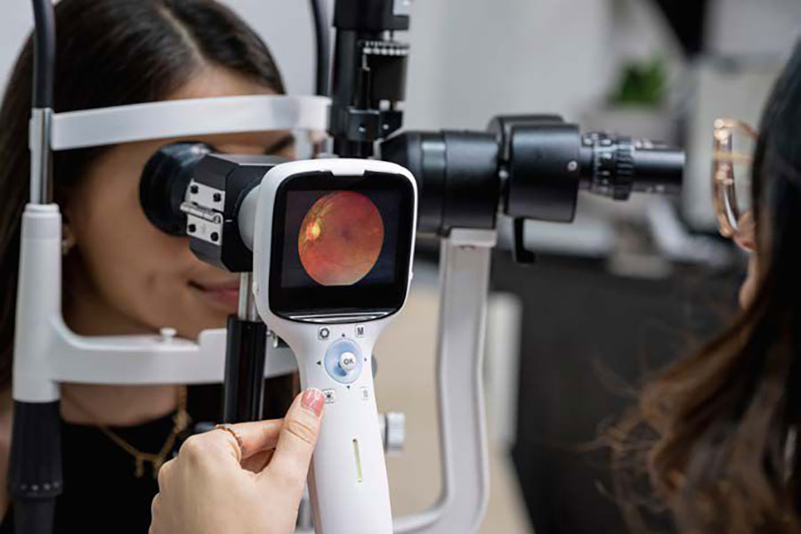
First, it started with fundus camera images where we could visualize and manipulate the retinal image to better understand what we are seeing under the microscope. Soon that evolved into wide angle retinal imaging to be able to get into the mid and far peripheral retina through an undilated pupil. This game changing technology allowed for a more expedient diagnosis (I sometimes make a retinal referral while the patient is in pre-testing and the image shows an obvious retinal detachment). Next came OCT (Ocular Coherence Tomography) which is essentially allowing a cross-sectional look under the retina and between retinal layers. OCT suddenly gave optometry the ability to instantly diagnose many more retinal pathologies with a high degree of certainty.
When we first acquired these technologies, we imaged every patient to better understand what we were seeing. The amount of occult pathology that was present without obvious symptomology made us pause and think.
Finally, we decided on a wellness model. We would image all patients with angle retinal imaging and OCT to screen for all pathologies that would present with symptoms and those that would be lurking as threats in the future.
Glaucoma Screening
Glaucoma is often called the silent thief of sight because there are often no obvious symptoms until vision is lost and major irreversible damage has taken place. Every eye exam looks for glaucoma, usually through intraocular pressure measurements and viewing the health of the optic nerves.
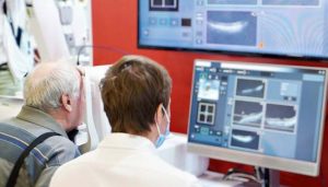
Nerve Fiber Layer (NFL) and Ganglion Cell Complex (GCC) imaging has allowed a look at early cellular changes before functional vision loss takes place. We screen all patients with visual fields, NFL and GCC scanning to set baselines for future reference and to identify glaucomas early in order to intervene and protect the future of our patients’ vision.
Dry Eye Disease
When I started practice, dry eye disease was a “grin and bear it” condition. Practitioners would acknowledge the presence of inflammation and provide artificial tears (usually a sample not a therapeutic course) and not follow up with the patient again until their routine eye examination two years later.
Now we screen for dry eye conditions before the patient arrives with a DEQ 5 dry eye survey and have added lower lid meibography and vital dye staining to every primary eye examination. Treatment advances in Dry Eye therapies abound and include advanced treatments such as Radio Frequency and IPL as well as LipiFlow. Medications that promote corneal healing and better tear production have made significant improvements in the lives of my patients previously suffering through untreated ocular surface disease.
Neurolens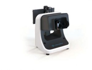
Recently we added an instrument that measures binocular dysfunction in a very accurate way through the Neurolens Measurement Device, or NMD. This technician-driven diagnostic test provides repeatable measurements that allow optometrists to prescribe Neurolenses, which incorporate a “contoured prism” to relieve symptoms associated with a proprioceptive conflict between misaligned eyes and the trigeminal nerve—sometimes referred to as trigeminal dysphoria. These common symptoms include dry eye sensation, light sensitivity, headaches and fatigue.
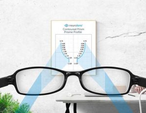
The results so far have been outstanding. Previously we would have to have patients suffer from not loving their progressive lenses and having to just “deal with it”. Patients have been raving! One patient described these glasses as “magic” and having changed her life.
It is rare that in one’s job you can make such a difference so as to evoke an emotional response of tears of joy and gratitude. Neurolens is such a technology, and a new way of prescribing glasses. We have started to screen all patients with symptoms of trigeminal dysphoria as Neurolens moves from being a problem solver to a
problem predictor.
The Rate Change Paradox
They say that change has never been this fast before and will never be this slow again. The rate change paradox requires optometric clinicians to change, learn and improve. As independent clinics, we have the power to instantly change without the required green light from head office that chains or corporate entities may require.
This is both a blessing and a curse. The curse is that nobody is requiring you to change via a memo sent down from the suits in HQ. That fact means that as leaders and practice owners the impetus for change has to come from you!
Embrace it! There will be many advances that will help us make the vision and indeed the lives of our patients more enjoyable. How quickly will you incorporate this into your clinic? The best is yet to come!

DR. TREVOR MIRANDA
Dr. Miranda is a partner in a multi-doctor, five-location practice on Vancouver Island.
He is a strong advocate for true Independent Optometry.
As a serial entrepreneur, Trevor is constantly testing different patient care and business models at his various locations. Many of these have turned out to be quite successful, to the point where many of his colleagues have adopted them into their own practices. His latest project is the Optometry Unleashed podcast.


